
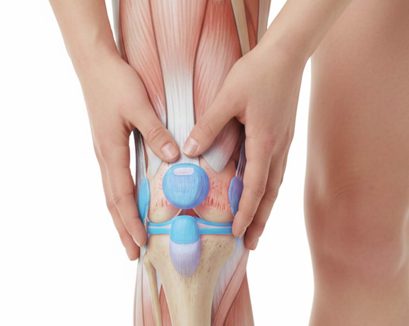
Why might I need an MSK ultrasound?
An MSK scan may be appropriate when pain, swelling, restricted movement or a soft-tissue injury needs closer assessment.
- Shoulder pain or reduced range of movement
- Knee pain or swelling
- Ankle pain after a sprain or twist
- Wrist, hand or elbow pain
- Hip-region pain or stiffness
- A lump or swelling near a joint
- Persistent symptoms after a sports injury or fall
- Suspected tendon or selected ligament injury
- Joint stiffness that has not improved with rest
What can an MSK ultrasound show?
What can be seen depends on the body area, the depth of the structure and the clinical question.
- Tendon inflammation, degeneration or tears
- Selected ligament sprains or tears
- Muscle tears and soft-tissue injury
- Joint effusion or fluid around a joint
- Bursitis
- Cysts or selected soft-tissue lumps
- Selected nerve compression, such as carpal tunnel syndrome
- Some features associated with inflammatory joint conditions
Conditions an MSK ultrasound may help assess
Depending on the area, ultrasound may help assess rotator-cuff tendinopathy or tears, tennis or golfer's elbow, Achilles tendinopathy, plantar-fascia problems, carpal tunnel syndrome, bursitis, muscle tears and selected ligament injuries.
A scan result is interpreted with your symptoms and clinical history. It may guide physiotherapy, GP review, specialist referral or a different form of imaging.
How does an MSK ultrasound work?
Gel is applied to the skin and a handheld transducer is moved over the area being examined. The sound-wave echoes are converted into real-time images on screen.
The clinician may compare positions or ask you to move during the scan. This dynamic assessment can show the movement of some tendons and soft-tissue structures.
What are the limitations of MSK ultrasound?
Ultrasound does not see through bone and may not fully assess structures inside a joint or deep within the body. MRI, X-ray, CT or specialist assessment may be more appropriate for some suspected fractures, bone conditions, cartilage injuries, meniscal problems or deeper structures.
The clinic should confirm whether ultrasound is suitable for the exact body area and symptoms before the appointment.
How to prepare for your MSK scan
No special preparation is usually required. Wear loose, comfortable clothing that allows easy access to the area being scanned and follow any instructions provided with the booking.
Bring previous imaging reports or relevant clinical letters if available, as these may help the clinician understand changes over time.
What to expect and how results are provided
The clinician will discuss the reason for the scan, explain the examination and answer questions. Appointments typically take 15 to 30 minutes depending on the body area and complexity.
Verbal feedback is discussed at the appointment, followed by a written report and images. The clinic will confirm the reporting timetable and any recommended next step when the booking is accepted.