Why might I need a gynaecological ultrasound?
A private gynaecological ultrasound may be considered when symptoms or fertility questions require assessment of the uterus, ovaries and other pelvic structures.
- Pelvic pain or discomfort
- Heavy, prolonged or irregular periods
- Bleeding between periods or after menopause
- Bloating or a feeling of pelvic pressure
- Suspected ovarian cysts or fibroids
- Irregular or absent periods
- Difficulty conceiving and seeking non-pregnancy fertility investigation
- A need to track ovulation and follicle growth
- Questions about ovarian reserve or egg count
- Symptoms associated with PCOS, such as irregular cycles or acne
What can the ultrasound show?
The structures assessed depend on the reason for the scan and the examination selected.
- Uterus size, shape and lining thickness
- The presence of fibroids or polyps
- Ovarian structure, cysts or follicles
- Follicle development and ovulation timing
- Antral follicle count as part of ovarian-reserve assessment
- Features associated with polycystic ovaries
- General pelvic anatomy relevant to the symptoms described
What conditions can a gynaecological ultrasound help identify?
A gynaecological ultrasound can help identify findings associated with ovarian cysts, uterine fibroids, polycystic ovaries, endometrial thickening and irregular ovulation patterns.
Ultrasound findings need to be interpreted alongside your symptoms and clinical history. Further investigation or specialist review may be recommended.
How does a gynaecological ultrasound work?
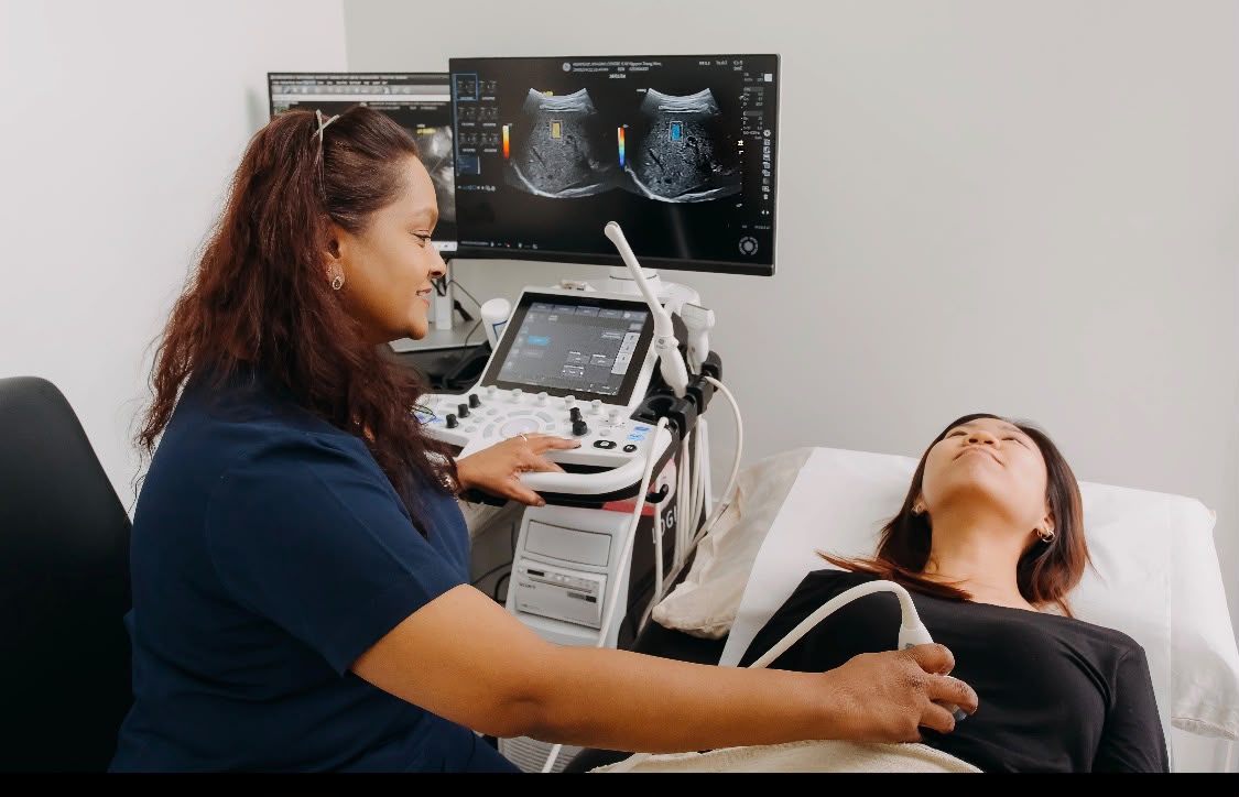
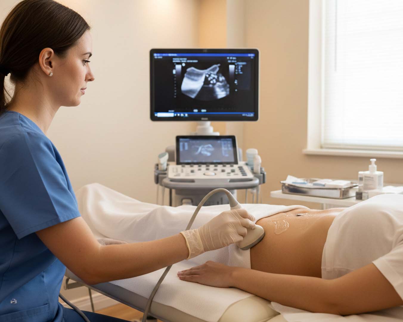
For an abdominal pelvic scan, gel is applied to the abdomen and a handheld transducer is moved over the skin. A transvaginal probe may be recommended when closer views of the ovaries, womb and uterine lining are needed.
The probe sends and receives sound waves to create real-time images. The examination type will be explained beforehand, and you can ask questions before giving consent.
Is a gynaecological ultrasound safe?
Diagnostic ultrasound uses sound waves rather than ionising radiation and is considered a low-risk imaging method. It can be used repeatedly when clinically appropriate, including for follicle tracking across a menstrual cycle.
Some people experience temporary pressure or mild discomfort from a full bladder or an internal probe. Tell the clinician immediately if you are uncomfortable.
How to prepare for your scan
An abdominal pelvic scan may require a full bladder to improve image clarity. A transvaginal examination normally requires an empty bladder.
If the appointment is for follicle tracking, provide the date of your last period when booking so the clinic can advise on timing. Always follow the preparation instructions supplied with your appointment.
What to expect on the day
The sonographer will explain the procedure, confirm the reason for the examination and answer questions before the scan begins. Appointments typically take 15 to 30 minutes.
You should be able to return to normal activities immediately afterwards unless the clinician advises otherwise.
Getting your results
Verbal feedback is discussed at the appointment, followed by a written report and images. The clinic will confirm the exact reporting arrangement when the booking is accepted.
If further investigation, referral or specialist input is recommended, the clinician will explain the appropriate next step.